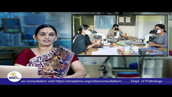
Overview
The Department of Pathology plays a fundamental role in patient care, and impacts nearly all aspects of patient care, from diagnosing cancer to managing chronic diseases through accurate laboratory testing.


Equipped with latest instruments like Fully automated IHC/ISH System and Fully automatic tissue processors.
A. ROUTINE TESTS:
1. Histopathological examination of small, medium and large biopsy specimens.
2. Intraoperative Frozen Sections and Stereotactic biopsy done on cryostat machine.
3. Decalcification and processing for bone biopsies.
B. SPECIAL TESTS:
1. Wide range of Histochemistryspecial stains.
2. Immunohistochemistry- Using a large panel of about 100 antibodies we are providing comprehensive immunohistochemistry to aid our histopathological diagnosis & providing therapeutic & prognostic significance for the patient management.
3. Immunofluorescence – Diagnostic aid in renal and dermatological pathologies.
A. ROUTINE TESTS:
1. Complete Hemogram, Reticulocyte count done in automated blood cell counters.
2. ESR, Sickling test, Absolute eosinophil count,Peripheral smear study.
3. Coagulation studies like Prothrombin time (PT)/ Activated partial thromboplastin time (APTT), Fibrinogen, Mixing studiesall done in fully automated coagulation analyzers.
4. Demonstration of LE cell and hemoparasites: Malarial Parasite, microfilaria.
5. Rapid screening tests for Malaria and Dengue.
B. SPECIAL TESTS:
1. Bone marrow aspiration/ trephine biopsy with cytochemistry.
2. Osmotic fragility test.
3. Flow cytometry - Used for diagnosis of Leukemia, enumeration of CD34+ hematopoietic stem cells (HSC) as a reference point for undertaking apheresis and evaluation of adequacy for peripheral blood stem cell (PBSC) engraftment, CD 19/20 count and CD4/CD8 enumeration.
TESTS DONE:
1. Urine routine/microscopy.
2. Urine RBC cytology, eosinophils, BJP, hemosiderin.
3. Stool, occult blood, fat, pH, reducing sugar.
4. Semen analysis.
5. Body fluids-cell count, crystals.
TESTS DONE:
1. FNAC.
2. Exfoliative cytology (conventional and liquid based cytology) including PAP smears, body fluids, sputum, bladder and bronchial brushings.
3. Cell block preparations for fluids and aspirates.
Pathology department at Amala helps doctors understand diseases by examining tissue and cell samples. A pathology laboratory in Thrissur plays an important role in diagnosing conditions ranging from inflammation and infections to certain cancers.
A histopathology laboratory examines tissue samples under a microscope to look for abnormal changes. These tests help doctors understand the nature of a growth, inflammation, or other changes found during an examination or procedure.
When in need to look at an unusual growth, lump or tissue change, a biopsy will be recommended. To help derive a diagnosis, a small sample is collected.
In most cases, yes. You can understand if a growth is benign or malignant by examining the tissue. But to find out the exact type of disease, some additional test might also be needed. You can visit the best biopsy testing laboratory in Thrissur – AIMS to get the most accurate diagnosis.
We can understand and get information about the type and features of a tumour through Pathology reports. These details help cancer specialists decide which treatments may be most appropriate and whether further tests are needed.
Not necessarily. Inflammation, infection, harmless growth or other conditions can cause abnormal cells as well. Only trust your doctor’s verdict rather than diagnosis or deriving answers yourself.
Some samples are straightforward to examine, while others require additional staining, testing, or review. More complex investigations naturally take longer, especially when a precise diagnosis is important for treatment planning.